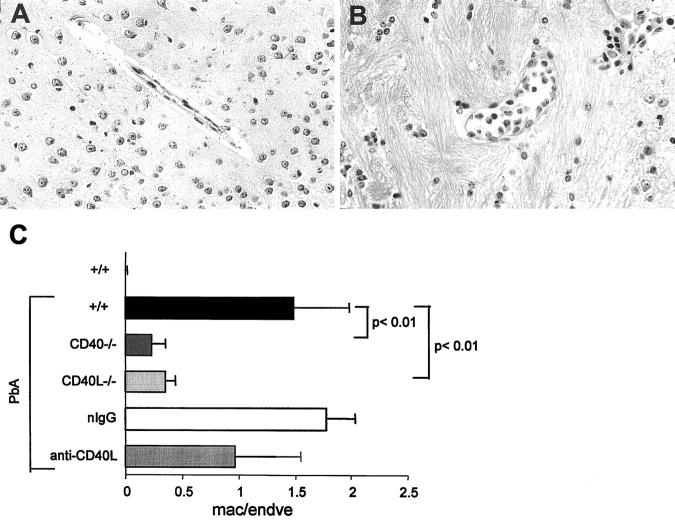
Figure 6.
Cell sequestration in brain venules. A and B: Cortical venules of infected +/+ (A) or CD40−/− (B) mice. An important sequestration of leukocytes is evident in A, but not in B (original magnification, ×400). C: Macrophages and endothelial cells in cortical venules were counted on H&E-stained sections by light microscopy. The results are expressed as the mean (+SD, n = 5) of the ratio of macrophages/endothelial cells seen in cortical venules.

