
Figure 7.
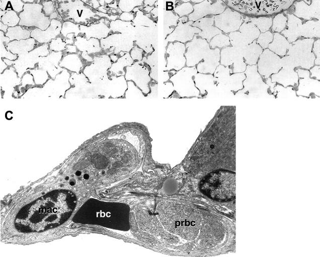
A and B: Alveolar capillaries from an infected +/+ (A) or CD40−/− (B) mice. An increased cellularity of the alveolar septa is evident in A but not in B, which is similar to noninfected +/+ mice (not shown). Macrophage sequestration is also evident in the venules (v) in A but not in B. C: An alveolar capillary from an infected +/+ is seen by transmission electron microscopy. A macrophage (mac), a red blood cell (rbc), and a parasitized red blood cell (prbc) in an advanced stage of maturation are seen inside the capillary. Original magnifications: ×400 (A and B), ×6500 (C).