
Figure 3.
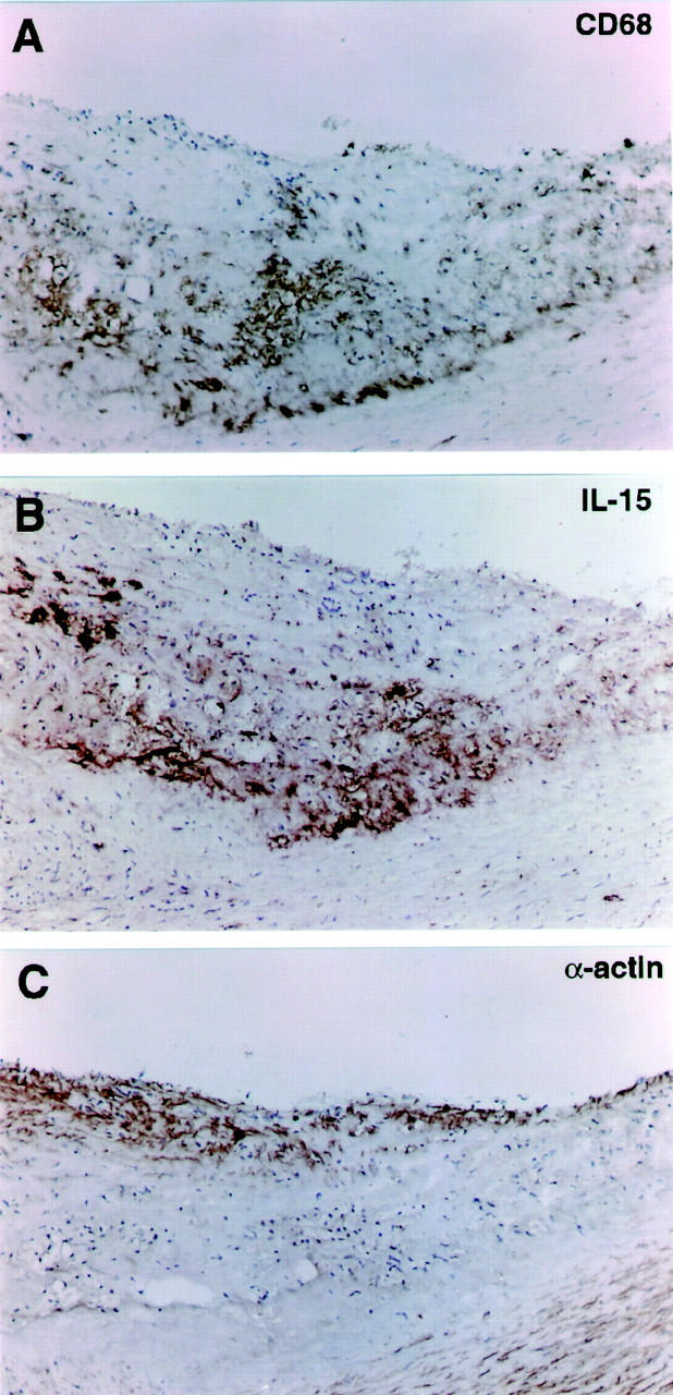
Immunohistochemical analysis of IL-15 in serial sections of human atherosclerotic lesions. Antibody binding was visualized by the avidin-biotin-peroxidase detection system. Original magnifications, ×100. A: CD68 staining for macrophages is most abundant in the core of the lesion. B: IL-15 staining co-localized with the macrophage-rich area in the core of the lesion. C: α-Smooth muscle actin staining depicts smooth muscle cells both in the media and in the cap of the atherosclerotic lesion.