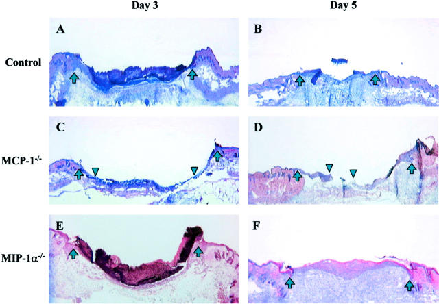
Figure 1.
Histological comparison of wounds from C57BL/6 controls (A and B), MCP-1−/− (C and D), and MIP-1α−/− (E and F) mice on day 3 (A, C, and E) and day 5 (B, D, and F) after injury. H&E-stained sections were photographed at ×25 power. The wound margins are indicated by upward arrows. In C and D, the margins of the advancing epithelial layer are indicted by downward arrowheads. By 3 days after injury, most wounds from control and MIP-1α−/− mice showed complete re-epithelialization (A and E). In contrast, day 3 wounds from MCP-1−/− mice showed delayed re-epithelialization (C). At day 5, wounds from MCP-1−/− mice still exhibited incomplete re-epithelialization (D).

