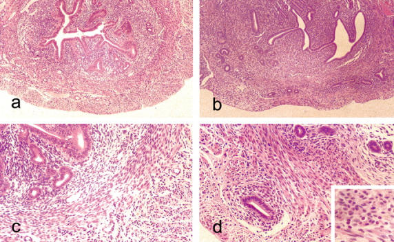
Figure 1.

Light microscopic appearances of the mouse uterus at 90 days. a: Untreated control showing endometrial glands and stroma surrounded by well-defined, concentric layers of smooth muscle cells. H&E: original magnification, ×55. b: Uterus from anti-estrogen (tamoxifen)-treated mouse showing adenomyosis with endometrial glands penetrating deeply into a myometrium that is devoid of the concentric bands of smooth muscle seen in the controls. H&E: original magnification, ×55. c: Higher power view of section seen in a showing well-defined layer of smooth muscle that sharply demarcates the endometrium. H&E: original magnification, ×140. d: Same case as in b showing glands penetrating the myometrium. The epithelium lining the penetrating glands shows little or no evidence of hyperplasia. H&E: original magnification, ×140. Inset shows the polymorphonuclear cells within the myometrium. H&E: original magnification, ×220.
