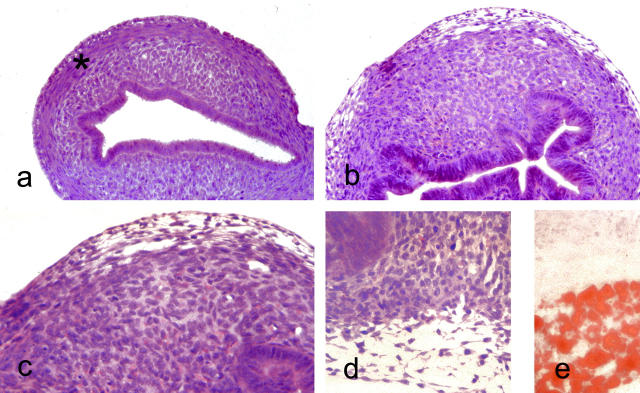
Figure 2.
Light microscopic appearances of uterus of 6-day-old mice. a: Untreated mouse uterus showing slit-like endometrial lumen lined by columnar cells with well-defined layers of endometrial stroma and smooth muscle. H&E: original magnification, ×140. b: Anti-estrogen (toremifene)-treated mouse at 6 days showing slightly hyperplastic endometrial glands and complete disruption of the stroma that is composed of stroma-like tissue with prominent small blood vessel. H&E: original magnification, ×140. c: Higher power view of stroma from b. At the periphery of the uterus, a layer of clear cells is evident. H&E: original magnification, ×220. d: The vacuolated nature of the clear cells, mature forms of which contained lipid (e: Oil red O; original magnification, ×220).

