
Figure 5.
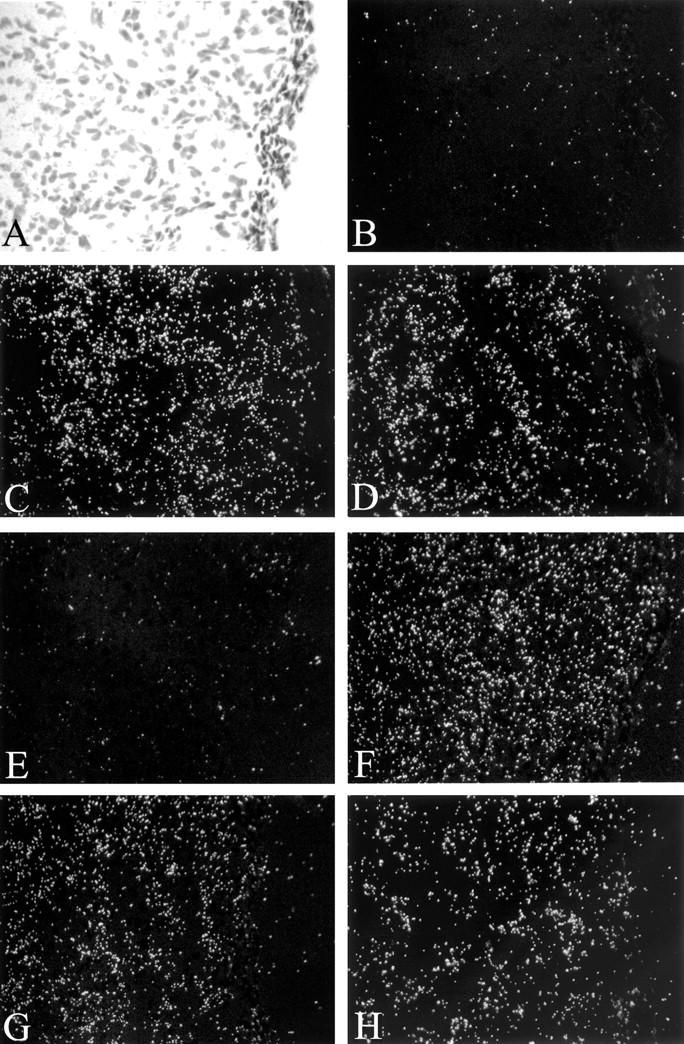
In situ hybridization of proliferative phase hemangioma. Bright-field photomicrograph (A) and dark-field photomicrographs of corresponding areas labeled by in situ hybridization for VEGF (B), Flt-1 (C), KDR (D), Ang1 (E), Ang2 (F), Tie1 (G), and Tie2 (H) mRNAs. Note strong expression of Flt-1, KDR, Ang2, Tie1, and Tie2 mRNAs, and in contrast weak expression of VEGF or Ang1. Original magnification, ×200.