Figure 3.
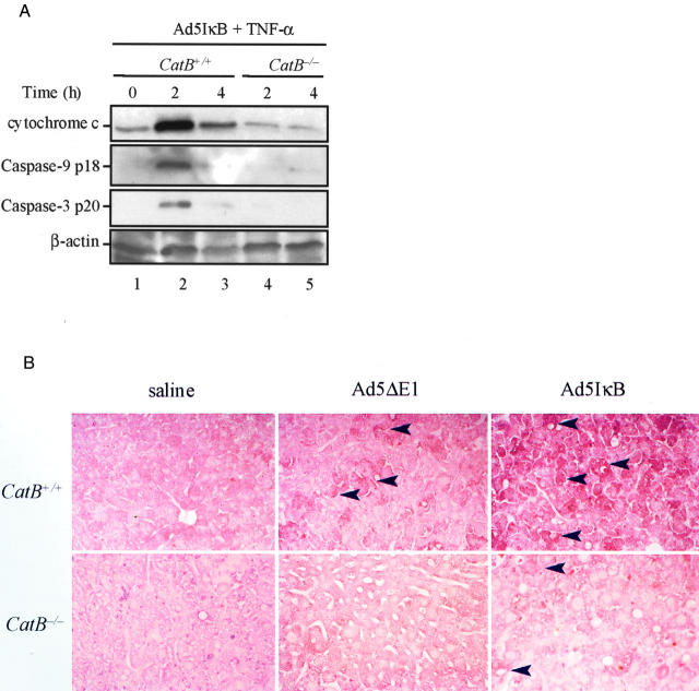
The mitochondrial pathway of apoptosis is activated in catB+/+ but not in catB−/− mice. CatB+/+ and catB−/− mice were injected with the adenovirus Ad5IκB. Twenty-four hours later, mice were treated intravenously with TNF-α and sacrificed after 2 and 4 hours. A: At the indicated time points, cytosolic fractions were prepared as described in Materials and Methods and aliquots of 60 μg of protein were subjected to sodium dodecyl sulfate-polyacrylamide gel electrophoresis on a 15% acrylamide gel, transferred to nitrocellulose membrane, and sequentially blotted for cytochrome c, active caspase 9, and active caspase 3. Cytosolic cytochrome c and active caspase 9 and 3 were only detected in catB+/+-treated livers. Blot for β-actin served as a control for protein loading. B: Representative immunohistochemistry for active caspase 3 and 7 in catB+/+ and cat B−/− mice injected with saline, Ad5ΔE1 (controls), or Ad5IκB, after a 2-hour treatment with TNF-α. Strongly positive immunochemical staining reaction for active caspase 3 and 7 was detectable in the cytoplasm of hepatocytes from Ad5IκB-injected catB+/+ mice after treatment with TNF-α, but not from catB−/− mice or in controls. The tissue sections were counterstained with eosin. Original magnifications, ×25.

