
Figure 2.
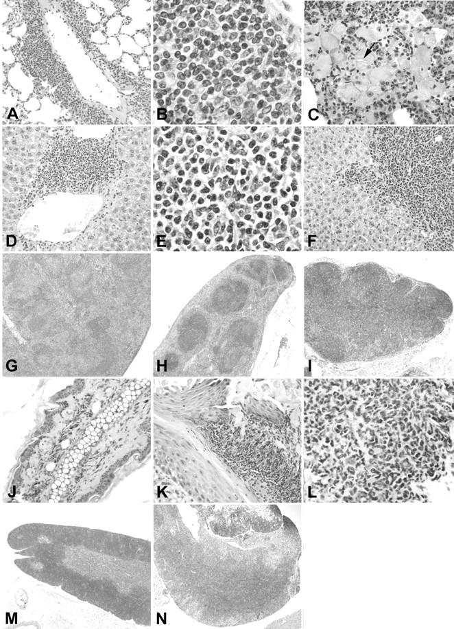
Systemic involvement in TSLP transgenic mice. A, B, and C: Lungs from TSLP transgenic mice. A perivascular leukocytic infiltrate (A) is present in the lung of a TSLP transgenic male without significant involvement of the alveoli. B: The higher magnification of A illustrates a mixed leukocyte population. C: Lung of a TSLP transgenic female at the age of 84 days. The alveoli are filled with large macrophages, some of which contain crystalline material (arrow). D, E, and F: Livers of TSLP transgenic mice. A mixed perivenous leukocyte infiltrate is present in D and E. Further spreading of the leukocyte infiltrate into the liver parenchyma is illustrated in F. G and H: Spleen of a TSLP transgenic male at the age of 111 days (G) and a wild-type control (H) at the same magnification. I: Enlarged mediastinal lymph node from a TSLP transgenic male at the age of 119 days. J: Ear of a wild-type control. K and L: Ear of a TSLP transgenic male at the age of 142 days demonstrates a prominent leukocyte infiltrate of the cutis. M: Thymus of a wild-type control. N: Illustrates the mediastinal tissue removed from a TSLP transgenic male at the age of 119 days. Original magnifications: ×40 (H, I, M, and N); ×200 (A, D, F, J, and K); ×400 (C and L); ×1000 (B and E).