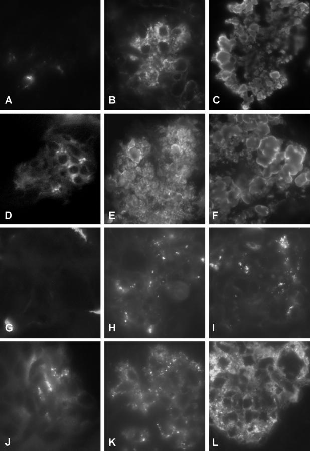
Figure 7.
Characterization of the immune deposits by immunofluorescence. A, D, G, and J: Renal specimens from a wild-type female stained for IgM (A), IgG (D), C3 (G), and IgA (J). The tissue shows weak and focal positivity for IgM and IgG, no glomerular C3 deposition, and positivity for IgA. B, E, H, and K: Renal specimen from a TSLP transgenic female stained for IgM (B), IgG (E), C3 (H), and IgA (K). Strong granular IgM and IgG deposits can be detected in the mesangium and in capillary walls. Focal C3 deposits can be seen in the same distribution. C, F, I, and L: Renal specimen from a TSLP transgenic male stained for IgM (C), IgG (F), C3 (I), and IgA (L).

