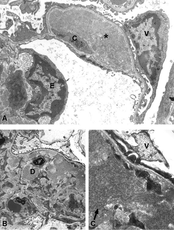
Figure 8.

Ultrastructual features of TSLP transgenic females. A: Transmission electron microscopy of a specimen from a 1-month-old TSLP transgenic female. The glomerular capillary shows a large subendothelial electron-dense deposit (asterisk), and cellular interposition (C). Note effacement of the foot processes over the deposit (V, visceral epithelial cell; E, endothelial cell). B and C: Transmission electron microscopy of a renal specimen from a 2-month-old TSLP transgenic female. Widening of the mesangial area because of increased matrix, and electron-dense deposits (D, deposit), with a tubular ultrastructure at higher magnification (arrow in C) (V, visceral epithelial cell). Original magnifications: ×4400 (A and B); ×16,900 (C).
