
Figure 1.
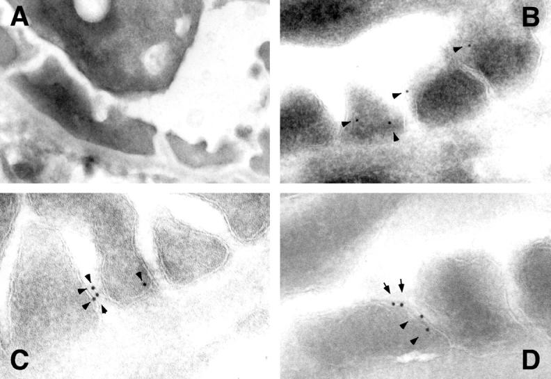
Immunoelectron microscopic localization of CD2AP in mouse podocytes. Immunoelectron microsopic localization of CD2AP in a 6-week-old normal mouse was performed using affinity purified CD2AP and secondary antibody conjugated with 10-nm gold particles. The immunogold particles (arrowheads) were found predominantly along the lateral borders of the podocyte foot processes (B–D). Many of the particles localize in a region near or at the slit diaphragm. Original magnification, ×20,000. No gold particles were seen in CD2AP KO kidney (A).