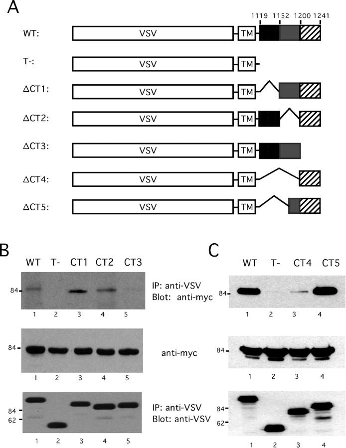
Figure 3.
Mapping the binding site for CD2AP in nephrin. A: Three cytoplasmic truncation mutants of nephrin, CT1 (lacking residues 1119–1151), CT2 (lacking residues 1152–1200), and CT3 (lacking residues 1201–1241), fused with VSV-G were generated to define the CD2AP binding site. B: Full-length myc-CD2AP and nephrin wild-type (WT), tail-truncated (T-), or cytoplasm-truncated mutants, were cotransfected as indicated in HeLa cells. VSV-G fusion proteins were immunoprecipitated with anti-VSV monoclonal antibody and immunoprecipitates were subjected to immunoblotting with anti-Myc antibody to detect CD2AP (top). The expression levels of VSV G fusion proteins were determined by re-probing the same blot using anti-VSV polyclonal sera. The expression levels of myc-CD2AP were determined by immunoblotting a sample of the cell lysate with anti-myc.

