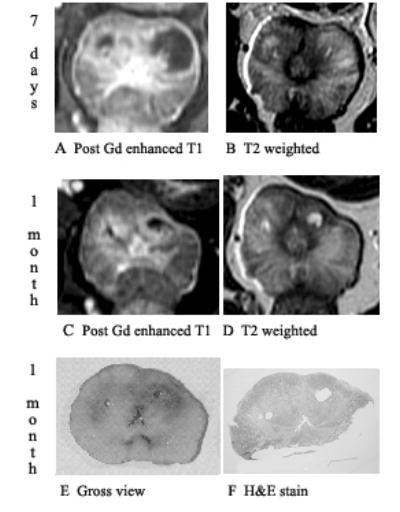
Fig. 5.

Prostate 7 days and 1 month after Tookad-PDT showing shrinkage of changes on post-gadolinium-enhanced T1-weighted images and signal changes on T2. Dog #5 (the left lobe received 150 J/cm and the right lobe 100 J/cm). Images are displayed with the dog’s anatomic left on the right side of the image. A: Post-gadolinium-enhanced T1-weighted images of 7 days post-PDT shows necrotic region well. B: On T2-weighted images of 7 days post-PDT, the margin of necrosis is poorly defined. C: Shrinkage of treatment effect on post-gadolinium-enhanced T1-weighted images at 1 month post-PDT. D: On T2-weighted images of 1 month post-PDT, cystic areas becomes very well defined. E: Gross view of whole mount. F: Pathologic slide shows two cystic areas surrounded by fibrosis corresponding to the unenhanced T1 and focal bright T2 areas.
