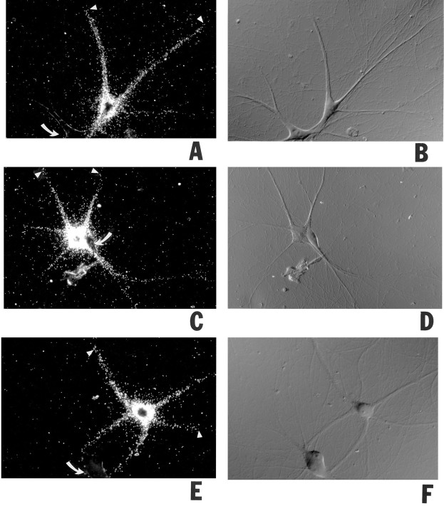
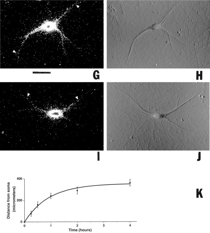
Fig. 5.
Progression of dendritic transport of injected BC1 RNA. Somata of sympathetic neurons in culture were microinjected with35S-labeled full-length BC1 RNA and were incubated at 35°C for 4 hr (A, B), 2 hr (C, D), 1 hr (E,F), 30 min (G,H), and 15 min (I,J), respectively. Arrowheadsindicate the distal-most points in dendrites at which BC1 labeling was detectable for a given time point. Curved arrowsindicate noninjected cells. A, C,E, G, I, Dark-field optics; B, D, F,H, J, Nomarski (DIC) optics. Scale bar, 100 μm. The measured average distances that the labeling signal had reached at each time point were as follows (mean ± SD in μm; sample sizes in parentheses): 15 min, 77 ± 24 (44); 30 min, 159 ± 28 (30); 1 hr, 242 ± 25 (45); 2 hr, 290 ± 33 (21); 4 hr, 360 ± 31 (24). The measured values were not dependent on injection amounts. The data are plotted inK (means, full circles; SD, error bars) with an exponential function (see Materials and Methods) to fit the data.


