
Fig. 1. Mesodermal-specific deletion of Tbx1 by Mesp1Cre.
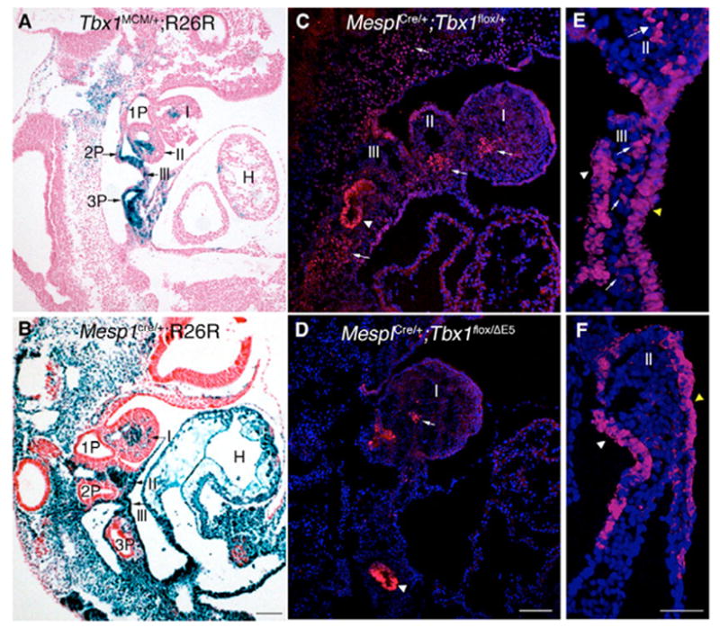
(A–D) Sagittal sections of E9.5 embryos. (A) Distribution of Tbx1-traced cells visualized by crossing Tbx1mcm/+ mice with the reporter R26R, compared with the distribution of Mesp1Cre-traced cells (B). Recombination is absent in the endoderm in the latter experiment. (C,D) Immunofluorescent staining with an anti-Tbx1 antibody on control (C) and conditional mutant (D) E9.5 embryos. Tbx1 immunoreactivity is preserved in the endoderm (arrowheads) and, to a lesser extent, in the core of the 1st pharyngeal arch (arrow in D), but not in other mesodermal domains (arrows). (E,F) Coronal sections of E9 embryos. Immunofluorescent staining with an anti-Tbx1 antibody on control (E) and M-ko embryos (F). Mesodermal expression (arrows) is eliminated in M-ko embryos, but endodermal (white arrowhead) and ectodermal (yellow arrowhead) expression are maintained. I, II and III, 1st, 2nd and 3rd pharyngeal arches; 1P, 2P and 3P, 1st, 2nd and 3rd pharyngeal pouches. Scale bar: 100 μm in A–D; 50 μm in E,F.