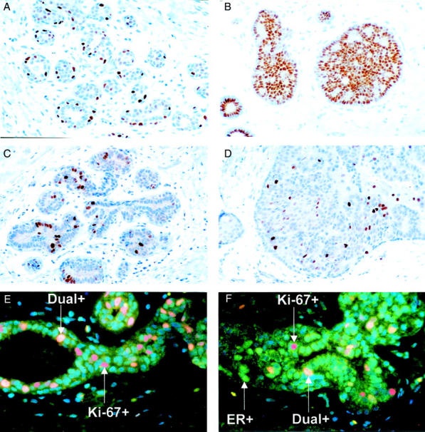
Figure 1.

A: ER-α expression. A normal breast lobule showing ER-α(+) cells surrounded by many ER-α(−) cells. B: ER-α expression. HUT biopsy from a case that subsequently developed breast cancer showing large numbers of ER-α(+) cells. C: Ki-67 expression. A normal breast lobule from a patient that progressed to breast cancer showing some Ki-67(+) cells among a majority of nonproliferating cells. D: Ki-67 expression. HUT focus from a case that subsequently developed breast cancer showing high proliferation rate. E: Indirect immunofluorescence for ER-α (green), Ki-67 (red), and dual-labeled cells (yellow). A normal breast lobule. F: Indirect immunofluorescence for ER-α (green), Ki-67 (red), and dual-labeled cells (yellow). HUT focus. Original magnifications: ×25 (A, B, C, and D); ×40 (E and F).
