
Figure 2.
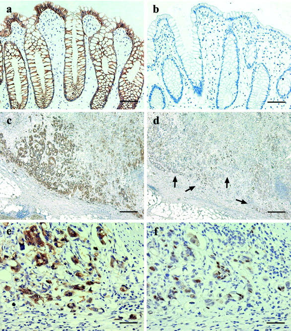
Sections of nonneoplastic colorectal mucosa (a and b) and colorectal carcinoma (c to f) immunostained for β-catenin (a, c, and e) and cyclin D1 (b, d, and f). The colorectal epithelial cells show strong membranous expression of β-catenin (a) and no detectable expression of cyclin D1 (b). The invasive edge of the carcinoma shows particularly prominent cytoplasmic and nuclear expression of β-catenin (c) and nuclear overexpression of cyclin D1 (d, arrows). Higher power views of the carcinoma show diffuse but heterogeneous nuclear expression of both proteins (e and f). The carcinoma cells also show prominent cytoplasmic β-catenin expression and loss of membranous expression (e). Scale bars: 150 μm (a and b); 600 μm (c and d); 75 μm (e and f).