
Figure 2.
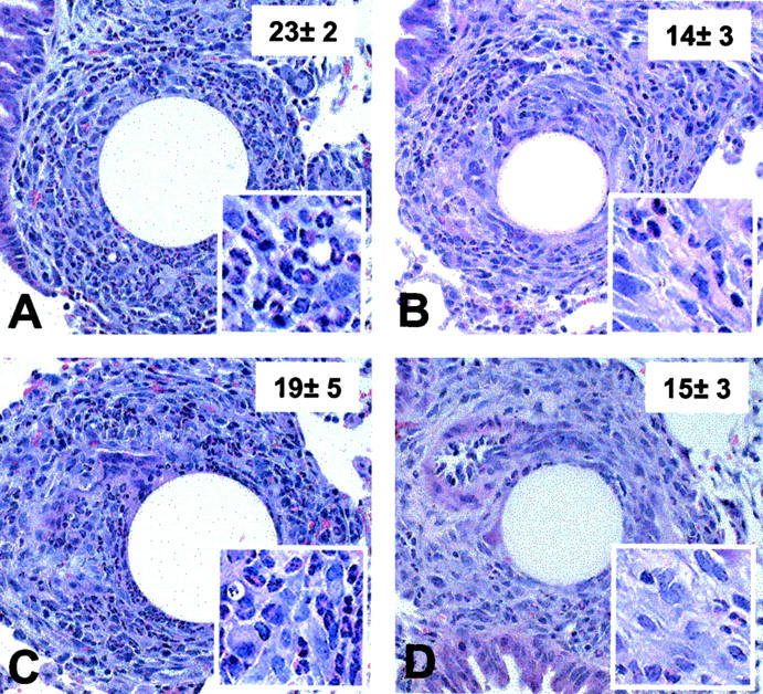
Histological appearance of type-2 (SEA) bead granulomas in mice treated with antibodies to eotaxin and MCP-3. Type-2 (SEA) lesions were generated in presensitized CBA mice, then at the time of bead challenge they were administered 10 mg of purified IgG (nonimmune rabbit, anti-eotaxin, anti-MCP-3, or 10 mg each of anti-eotaxin plus anti-MCP-3 Abs). On day 4 after Ab treatment and bead challenge, lung tissues were harvested and histological sections were prepared as described in Materials and Methods. A, Nonimmune IgG treated; B, anti-MCP-3 treated; C, anti-eotaxin treated; D, combined anti-MCP-3 and anti-eotaxin treatment. Granulomas from each of the five to six mice per treatment group were analyzed morphometrically and the inset numbers indicate mean eosinophil counts per 50 × 50 μm area ± SD. Four such areas were measured from each granuloma. Original magnifications: ×200 (A to D); ×400 (insets).