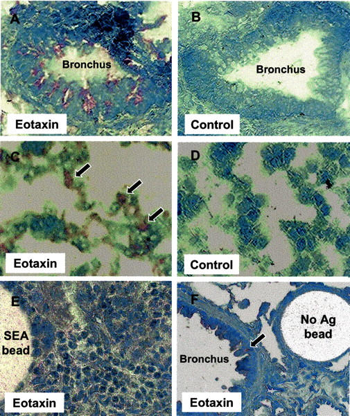
Figure 5.

Immunohistochemical localization of eotaxin. Frozen sections of lungs were prepared and stained with anti-eotaxin Abs as described in Materials and Methods. A: Bronchus from lung with type-2 (SEA) granulomas, eotaxin stain. B: Serial section of A, nonimmune Ab control. C: Lung parenchymal alveoli in lung with type-2 (SEA) granulomas, eotaxin stain. D: Lung parenchymal alveoli in lung with type-2 (SEA) granulomas, nonimmune Ab control; E, Type-2 (SEA) bead granuloma, eotaxin stain. F: Bronchus and nearby control bead lesion in lung challenged with antigen-free beads. Original magnifications: ×200 (A, C, F); ×400 (D, E).
