
Figure 10.
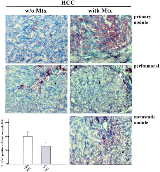
α3-integrin expression in tumoral, peritumoral, and metastatic HCC tissue. α3-integrin is localized in the parenchyma of metastatic (28 patients) and nonmetastatic (12 patients) HCC primary nodules (white arrows) with a similar distribution pattern, but is more strongly expressed in the metastatic tissue. In the peritumoral tissue of HCC patients with and without metastasis, α3-integrin is expressed by the blood vessel endothelial cells present in the stroma (white arrows) but is completely absent in the parenchyma (black arrows). In the metastatic nodule, α3-integrin is distributed as in the primary nodule. In the metastatic tissue, α3-integrin is expressed to the same extent as in the HCC primary nodule. α3-integrin expression level is higher in patients with metastasis than in those without (P < 0.001). Scale bar, 5 μm.