
Figure 5.
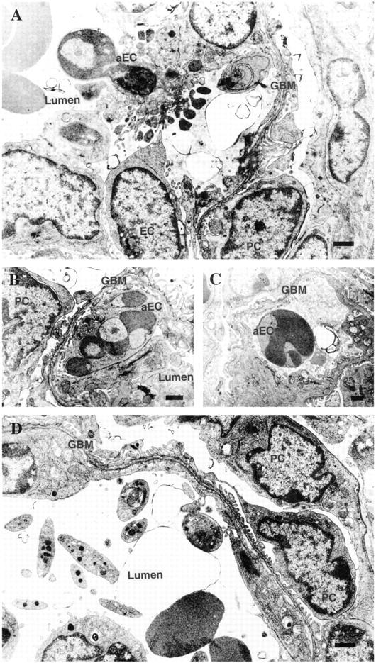
Electron microphotographs of kidney glomeruli from the mice injected with APB5 for 3 days (A–C) or 9 days (D). A: An apoptotic glomerular endothelial cell with many buddings and apoptotic bodies. B: An apoptotic glomerular endothelial cell containing fragmented nuclei with peripherally condensed chromatin. C: An apoptotic cell seen in the glomerular capillary lumen. D: At 9 dpp, glomerular capillary tufts were almost destroyed and blood cells (red blood cells, leukocytes, and platelets) were observed inside, presumably reflecting glomerular endothelial cell loss via apoptosis at younger ages. aEC, apoptotic endothelial cell; EC, glomerular endothelial cell; GBM, glomerular basement membrane; PC, podocyte. Scale bars, 1 μm.