
Figure 1.
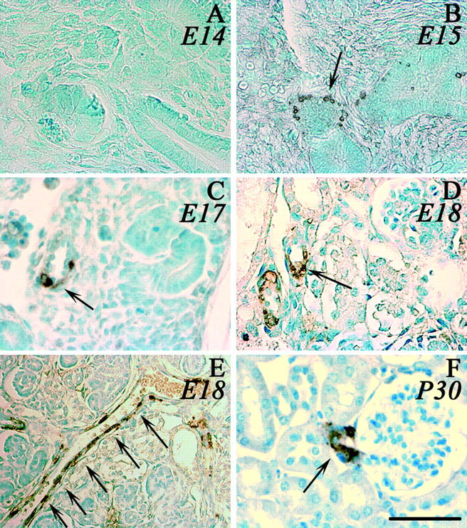
Renin expression during ontogenesis. Renin expression in the kidney of C57Bl/6 mice was detected by immunohistochemistry using the polyclonal anti-renin antibody CAS 16 detected by peroxidase staining. A–E: Photomicrographs of fetuses at different stages (A, E14; B, E15; C, E17; D, E, E18). E is photomicrograph from an interlobular artery in embryonic kidney (E18) and F from an adult kidney (P30). Arrows indicate examples of renin-expressing cells. Scale bar, 50 μm.