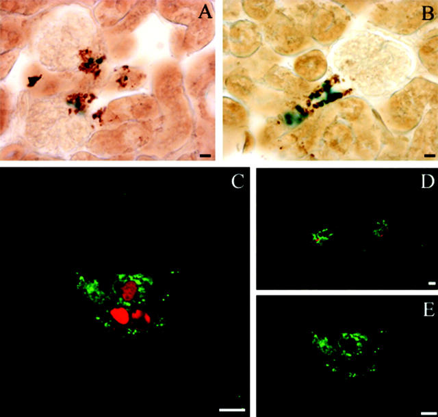
Figure 5.
Colocalization of β-galactosidase and renin expression in juxtaglomerular cells. β-Galactosidase and renin expression were detected in kidney sections by light microscopy (A, B) or confocal microscopy (C, D, E). A and B: β-Galactosidase (blue) expression was detected by X-gal staining in the nucleus of cells expressing endogenous renin (brown) in the cytoplasm. Renin expression localization was determined by immunohistochemistry with the CAS antibody 16 detected by peroxidase staining. C and D: Projection of 8 stacked images obtained by sequential scan acquisition. A similar colocalization was observed with a nuclear localization of β-galactosidase (red) in renin-expressing cells (green). E: Typical renin granules within the JG cells are shown in a confocal section with only FITC/renin fluorescence excitation. Scale bars, 10 μm.

