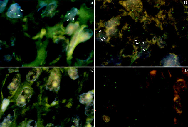
Figure 1.
Fluorescent in situ hybridization analysis of poorly differentiated (A, C) and anaplastic thyroid cancer (B, D). Spectrum red-labeled 13q12 BAC clone and spectrum green-labeled CEP 10 probes hybridized to the 5-μm section confirming decreased copy numbers in chromosome 13 identified by comparative genomic hybridization in these cases, as depicted by arrows identifying the signals (A, B). Spectrum red-labeled HER-2 probe and spectrum green-labeled CEP 17 probes hybridized to the 5-μm section confirming increased numbers in chromosome 17 identified by comparative genomic hybridization in these cases (C, D).

