
Figure 3.
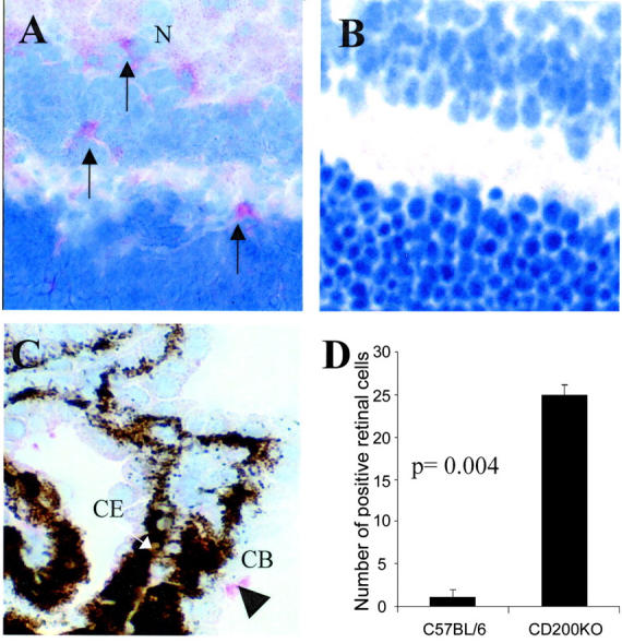
CD200−/− MG display increased constitutive NOS2 expression. Immunohistochemical sections demonstrating NOS2 expression detected with SA:ABC-AP. NOS2-positive MG cells were detected within normal CD200−/− retina (A), and primarily absent in CD200+/+ retina (B). C shows that within the CD200+/+ eye NOS2 expression was confined primarily to nonretinal sites such as ciliary body (CB) (arrowhead; CE, ciliary body epithelium that is pigmented). Counting confirmed that within the retina there is a significant increase in CD200−/− MG NOS2 expression. For quantification, mean NOS2-positive retinal cells were calculated from a minimum of three sections per eye at each time point (n = 9; groups of three, three sections per eye). Original magnifications: ×600 (A); ×650 (B); ×300 (C).