
Figure 5.
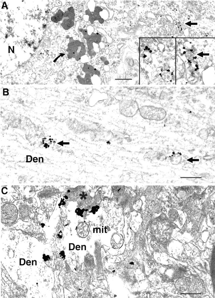
Aβ42 accumulates within neuronal soma and processes in human AD brain. A: Human AD cortical brain specimen showing substantial amounts of Aβ42 labeling within MVBs of an abnormal appearing neuronal soma (arrow); curved arrow indicates lipofuscin devoid of specific immunogold labeling. Nucleus has abnormal appearing accumulation of heterochromatin near the outer membrane. Insets represent higher power views of Aβ42 immunogold labeling of MVBs in neuronal soma. B: Multiple Aβ42 gold particles are associated with two MVBs in a dendrite. C: Multiple aggregated Aβ42 immunogold particles are found within a disrupted, swollen dendrite containing electron-dense material (asterisk), indicative of degeneration. Abbreviations: Den, dendrite; mit, mitochondrion; N, nucleus. Scale bars: 1 μm (A); 500 nm (B and C).