
Figure 5.
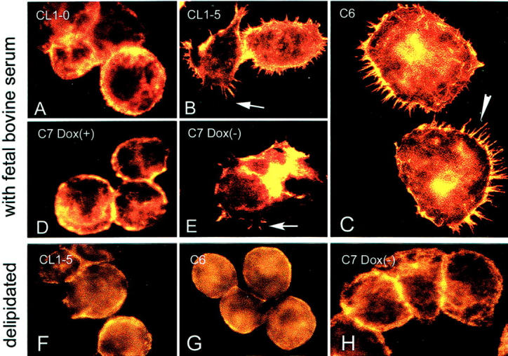
Caveolin-1+ve CL1-5 cells (B), C6 cells (C), and C7-Dox(−) cells (D) showed abundant filopodia formation. The caveolin-1−ve CL1-0 cells (the parental cells for C6, A) and C7-Dox(+) cells (E) did not reveal filopodia formation in cultures. However, the ability to form filopodia in CL1-5, C6, and C7-Dox(−) cells was abolished when cells were cultured in delipidated medium (F to H). A rhodamine phalloidin staining was used to highlight the presence of F-actin in filopodia and at the peripheral cytoplasmic area.