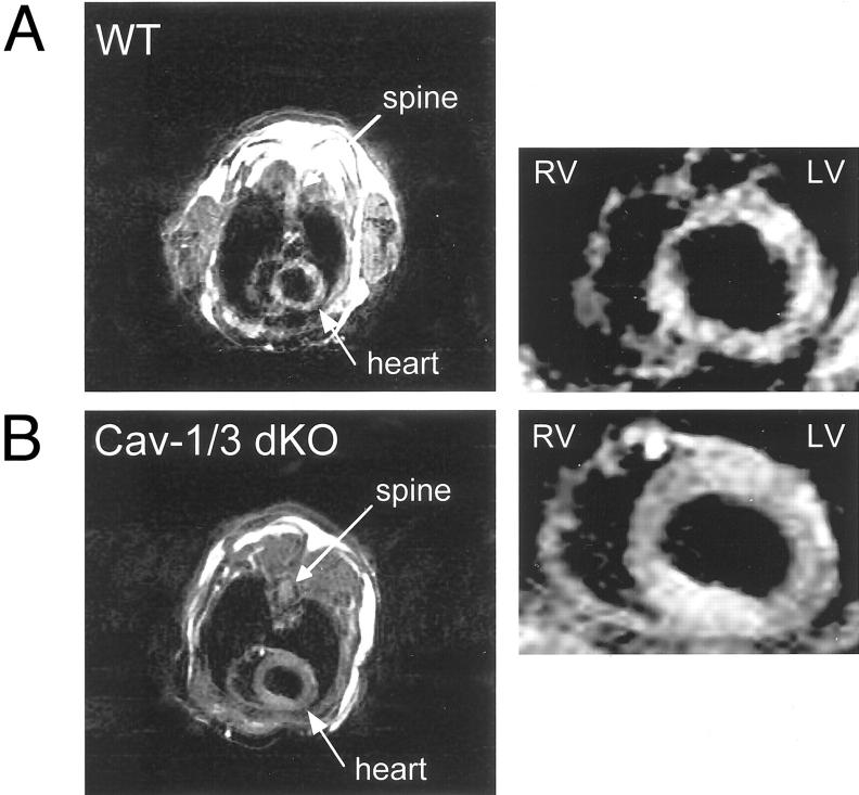
Figure 4.
Cav-1/3 dKO mice exhibit cardiac hypertrophy as assessed by gated MRI. Representative short axis (transverse) images at the mid-level of the heart of WT (A) and Cav-1/3 dKO (B) during diastole. Note the concentric hypertrophy of the left ventricle (LV) in the Cav-1/3 dKO heart. RV, right ventricle; LV, left ventricle.

