
Figure 1.
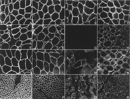
Integrin immunofluorescence studies. Transverse cryosections of muscle-biopsy specimen from a normal control and three patients with integrin deficiency were stained with antibodies directed against the intracellular domain of integrin α7B, integrin α7A, integrin β1D, and laminin α2. A–D show a normal muscle biopsy, E–H show a muscle biopsy showing complete integrin α7 deficiency, and I–R show muscle biopsies from two partial integrin α7-deficient patients. Integrin α7B immunostaining shows a uniform staining of the periphery of each myofiber in the control muscle (C), where a muscle biopsy showing a complete absence of immunoreactivity is shown in G. M: Integrin α7 is severely reduced. The majority of the muscle fibers show a barely detectable immunostaining, but a few, scattered fibers are strongly integrin α7-positive and show a slight cytoplasmic α7B-positive immunostaining. Q: A different pattern of integrin α7 partial deficiency is shown. Integrin α7-positive and -negative fibers are scattered in a mosaic-like pattern. The integrin-positive fibers show a α7B-positive cytoplasmic staining. Integrin α7A was reduced (H, N, R) and integrin β1D mildly reduced (F, L, P) in the complete integrin α7 deficiency muscle biopsy and in both the partial integrin α7 deficiency specimens in comparison with the control muscle (D and B). Laminin α2 was normal in all muscle biopsies (A, E, I, O).