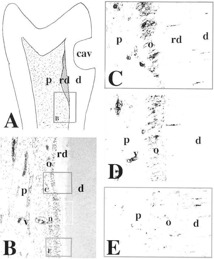
Figure 4.
Immunohistochemical localization of N-cadherin in sections of permanent human premolars after cavity preparation. A: Schematic illustration of a premolar showing reparative dentin production, 9 weeks after class V cavity preparation. The frame represents the area shown in the B. B: H&E staining. The frames indicate the areas shown in C, D, and E. C: A strong N-cadherin labeling is observed in odontoblasts producing the reparative dentin. Note that the staining is detected in both odontoblast bodies and odontoblast processes. D: N-cadherin reactivity is decreased in odontoblasts near to the injury site, but not involved in reparative dentin synthesis. Note that blood vessels are N-cadherin-positive. E: N-cadherin staining is very faint in odontoblasts far away from the injury site. Note that the staining persists in some of the odontoblast processes. Abbreviations: cav, cavity; d, dentin; o, odontoblasts; p, pulp; rd, reparative dentin; v, vessel.

