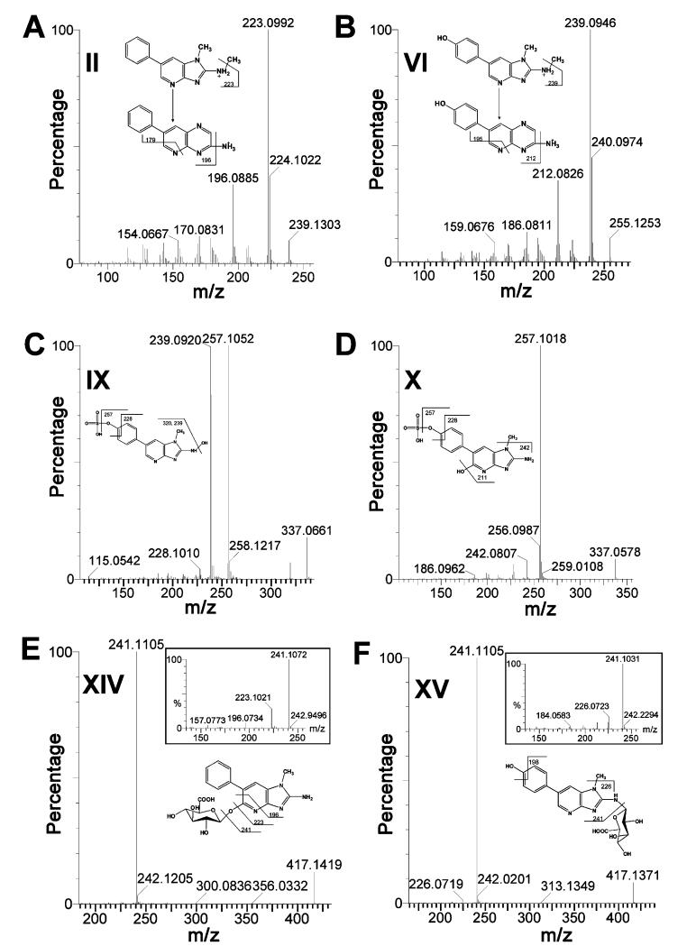
Figure 2.
LC-MS/MS structural elucidation of urinary PhIP metabolites. Conditions for LC-MS/MS were described in the Experimental Procedures. MS2 fragmentation was conducted with collision energy ramping from 15 to 35 eV. (A) MS2 fragmentation of N2-methyl-PhIP (II). (B) MS2 fragmentation of N2-methyl-PhIP-4′-OH-PhIP (VI). (C) MS2 fragmentation of N2,4′-diOH-PhIP sulfate (IX). (D) MS2 fragmentation of 5,4′-diOHPhIP sulfate (X). (E) MS2 fragmentation of 5-OH-PhIP glucuronide (XIV) with inlaid spectrum of deconjugated 5-OH-PhIP ion. (F) MS2 fragmentation of 4′-OH-PhIP-N2-glucuronide (XV) with inlaid spectrum of deconjugated 4′-OH-PhIP ion. Major daughter ions from fragmentation were interpreted in the inlaid structural diagrams.

