
Figure 6.
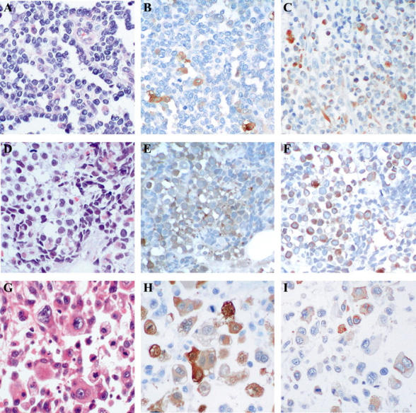
Arpp and Carp are strongly expressed in RMS cells. RMS tissues were analyzed by H&E staining (A, D, G) and immunohistochemistry with α-Arpp(FL) Ab (B, E, H) and α-Carp(N) Ab (C, F, I). In a case of alveolar-type RMS (A–C), Arpp-positive and -negative RMS cells (B) and Carp-positive and -negative RMS cells (C) are admixed. In a case of embryonal-type RMS (D–F), strongly immunoreactive RMS cells are scattered among those weakly stained (E). Population of Carp-positive RMS cells is larger than that of Arpp (F). Both nuclei and cytoplasm are positively stained (F). In a case of pleomorphic-type RMS (G–I), Arpp-positive and -negative RMS cells are scattered. Both cytoplasms and nuclei are positively immunostained (H). Carp-positive and -negative RMS cells are diffusely distributed (I). The population of Carp-positive RMS cells is lower than that of Arpp (I). Original magnifications, ×200.