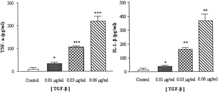
Figure 3.
Confluent brain endothelial cells were incubated in serum-free DMEM with 0.1% bovine serum albumin and then exposed to different concentrations of TGF-β for 24 hours. Control (unstimulated) cells were incubated in media alone. The supernatants were assayed for IL-1β (right) and TNF-α (left) using an ELISA-based assay. *, P < 0.05; **, P < 0.01;***, P < 0.005, significantly different from unstimulated endothelial cells.

