
Figure 1.
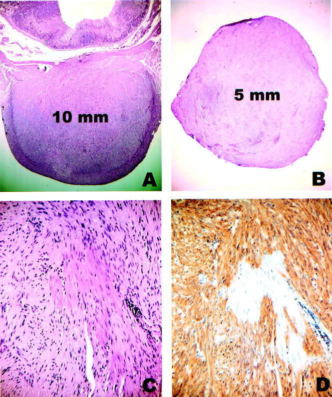
Incidental GISTs. A: H&E-stained section of case 6 showing a 10-mm tumor centered in the muscularis propria of the stomach. The lesion was discovered during a gastroesophagectomy for a large esophageal leiomyoma. B: H&E-stained section of case 11 showing a well-circumscribed 5-mm nodule that was removed from the serosal aspect of the small bowel during surgery for endometrial adenocarcinoma. C: Close-up of case 11. Note that entrapped smooth muscle cells of the muscularis propria are morphologically similar to the tumor cells. D: Same area of case 11 as in C, immunostained for KIT. The tumor cells are strongly positive whereas the smooth muscle cells are negative.