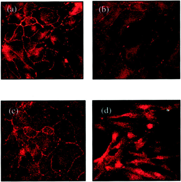
Figure 8.
Immunohistochemistry for occludin (a and b) and ZO-1 (c and d). Confluent monolayers of HK-2 cells were stimulated with TGF-β1 for 4 days before fixation and analysis by immunohistochemistry as described in Materials and Methods. Under control conditions (serum-free medium alone) both occludin (a) and ZO-1 (c) clearly outline the cell contour. After stimulation with recombinant TGF-β1, occludin staining intensity becomes weaker and discontinuous (b). ZO-1 staining after the addition TGF-β1 demonstrates relocation from the cell periphery into the cell cytoplasm (d).

