
Figure 5.
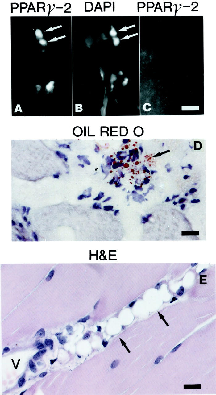
Early onset of adipogenesis in the perivascular space of ADAM 12 transgenic mice. A: Immunofluorescence staining using antibodies to PPARγ-2 on frozen sections of a 3-week-old ADAM 12-L transgenic mouse. Note the presence of scattered immunoreactive nuclei in the perivascular space. B: The same section was stained with 4,6-diamidino-2-phenylindole (DAPI) to stain the nuclei. C: Immunofluorescence staining using antibodies to PPARγ-2 on frozen sections of a 3-week-old littermate control mouse did not reveal any immunoreactive nuclei. D: Oil red O staining demonstrates small fat droplets in cells in frozen sections of a 3-week-old ADAM 12-L mouse. E: H&E-stained paraffin section of m. quadriceps femoris from a 3-week-old ASFM transgenic mouse shows several mature adipocytes. Arrows mark cells with PPARγ-2-immunoreactive nuclei and 4,6-diamidino-2-phenylindole-stained nuclei in A and B, respectively. Cells labeled by oil red O are marked with arrow in D. Mature adipocytes are marked with arrows in E. P, perivascular space; V, blood vessel. Scale bars: 60 μm (A–C); 40 μm (D and E).