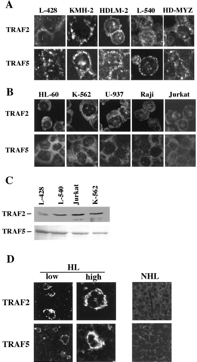
Figure 1.

A: Laser confocal immunofluorescence microscopy for intracellular distribution of TRAF2 and TRAF5 proteins. H-RS derived cell lines. Aggregation of TRAF2 and TRAF5 in the cytoplasm is clearly shown. Original magnification, ×400. B: Hematopoietic and lymphoid cell lines unrelated to Hodgkin’s disease. Both TRAF proteins are diffusely distributed in the cytoplasm. Original magnification, ×400. For A and B, following antibodies are used. First antibodies: anti-TRAF2 (C-20) rabbit antibody (Santa Cruz) and anti-TRAF5 (C-19) goat antibody. Secondary antibodies: FITC-labeled anti-rabbit donkey antibody (Amersham Pharmacia Biotech), FITC-labeled anti-goat donkey antibody (Santa Cruz). C: Immunoblot analysis of TRAF2 and TRAF5 showing little difference in the amounts of expressed TRAF proteins. D: Laser confocal immunofluorescence microscopy of biopsied lymph nodes for intracellular distribution of TRAF2 and TRAF5 proteins. HL, a lymph node of Hodgkin’s lymphoma; NHL, a lymph node of non-Hodgkin’s lymphoma (diffuse large B cell type). Instead of secondary antibodies, streptavidin-FITC or streptavidin-Texas Red was used in modified TSA system (NEN Life Science). Original magnification, ×200.
