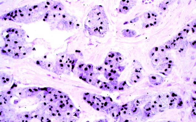
Figure 5.
Photomicrograph of a breast carcinoma displaying HER-2/neu gene amplification by FISH, mRNA enhanced expression, and overexpression of the oncoprotein by immunohistochemistry. Most of the nuclear area of the tumor cells contains large confluent black metallic gold signal generated through autometallography. GOLDFISH with nuclear fast red counterstain; original magnification, ×400.

