
Figure 1.
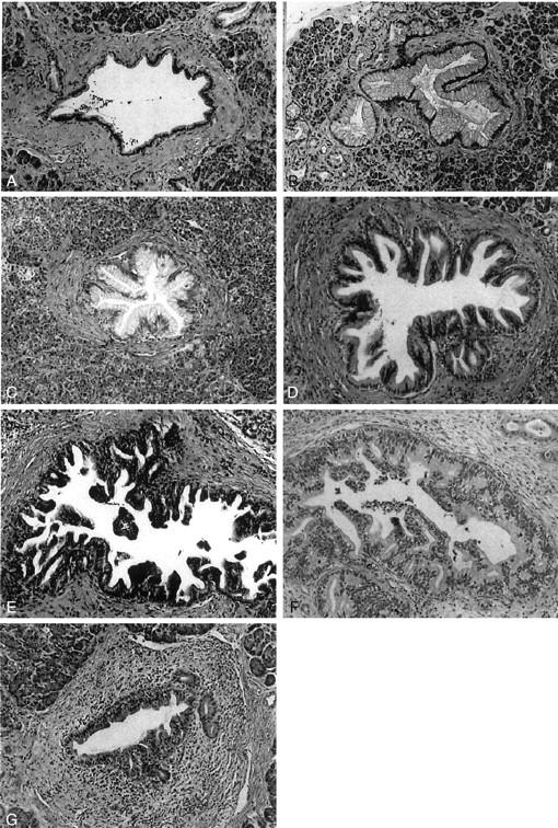
Pancreatic intraepithelial lesions. A: Normal pancreatic duct. The epithelium is composed of cuboidal to low columnar cells. B: PanIN-1A. The epithelial lesions are composed of tall columnar cells with abundant mucin in cytoplasm. The nuclei are small and located in the basal portion of the cells. C: PanIN-1B. It shows a papillary structure. The epithelial cells are similar to those in PanIN-1A. D: PanIN-2. The mucinous epithelial lesion shows mild to moderate irregularity of the papillary structure. Mild nuclear pseudostratification and enlargement (elongation) are seen. E: PanIN-3. Irregularity of the papillary structure is obvious. Budding-off of small clusters of epithelial cells into the lumen is seen. The epithelial cells show increased nuclear-to-cytoplasmic ratio, nuclear enlargement, and irregularity. F: Cancerization of ducts. This lesion is similar to PanIN-3. In surrounding stromal tissue, however, there is an infiltrating adenocarcinoma composed of cells similar to those in the intraductal lesion. G: Reactive change. There is inflammatory cell infiltration around the ductal lesion. Although mild to moderate nuclear enlargement is seen, other nuclear atypia such as hyperchromasia and irregularity of shape are not identified. H&E stain; original magnifications, ×100.