
Figure 2.
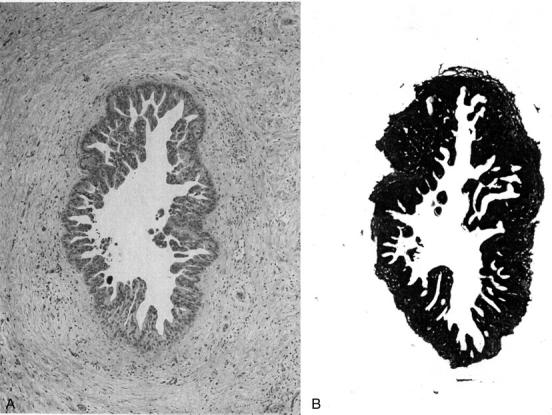
An example of microdissection. A: An intraductal epithelial lesion present in the histological section (H&E stain). B: Surrounding tissue was scratched and removed by the blade. The PanIN is easily collected by scratching the slide and aspirating the tissue after drops of TK buffer have been added.