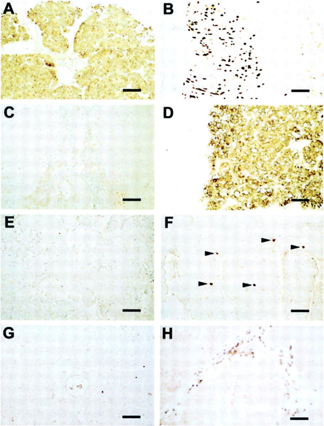
Figure 1.

Presence of HDC and absence of TH and TPH in SCLC tumors. Immunohistochemistry of a SCLC tumor (case 11 in Table 1 ▶ ), stomach, adrenal, and duodenum is shown. HDC immunostaining was found in the tumor (A) and in the ECL cells of the human stomach mucosa (B). No staining was observed in the SCLC tumor using antibodies directed against TH (C) and TPH (E), but strong immunostaining is apparent in adrenal medullary chromaffin cells (D) and in enterochromaffin cells (arrowheads) of the duodenum mucosa (F). Nonimmune rabbit serum (1:10,000) is shown as a negative control (G). The location of mast cells is shown by immunostaining with an antibody directed against tryptase. Groups of mast cells present in the stroma of the tumor are shown between the clusters of unstained SCLC cells (H). The size of the bars (equivalent to 100 μm) is given on the right.
