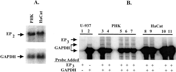
Figure 1.
EP2 receptor mRNA expression in PHKs and HaCat cells: A: Northern hybridization showing approximately equivalent expression of EP2 receptor in both nonconfluent PHKs and HaCat cells. Hybridization was done with a 32P-labeled EP2 receptor riboprobe (top) or housekeeping GAPDH riboprobe (bottom). Each lane represents 7 μg of poly(A)+-enriched RNA. B: RNase protection assay demonstrating decreased EP3 receptor transcript in HaCat cells compared with adult PHKs. Total RNA was prepared from positive control U-937 cells (lanes 1 and 2), adult PHKs from two separate individuals (individual I, lanes 3 and 4; individual II, lanes 5, 6, and 7), and HaCat cells (lanes 8 to 11). Lanes 3, 5, 8, and 10 represent total RNA isolated from nonconfluent cultures. Lanes 4, 7, 9, and 11 represent total RNA isolated from cultures at 3 to 4 days after confluence. Lane 6 represents RNA isolated from a culture that had just reached confluence. Cellular RNA (10 μg for lanes 1–7, 20 μg for lanes 8 and 9, or 40 μg for lanes 10 and 11) was incubated with a [32P-UTP]-labeled EP3 and/or a GAPDH riboprobe followed by RNase A/T1 digestion. Protected fragments were visualized using a phosphorimager after nondenaturing polyacrylamide electrophoresis as described in Materials and Methods. Lane 1 represents the protected EP3 fragment observed in positive control RNA hybridized with only the EP3 riboprobe. Lane 2 represents the protected fragments when only the GAPDH riboprobe is hybridized to control RNA.

