Figure 2.
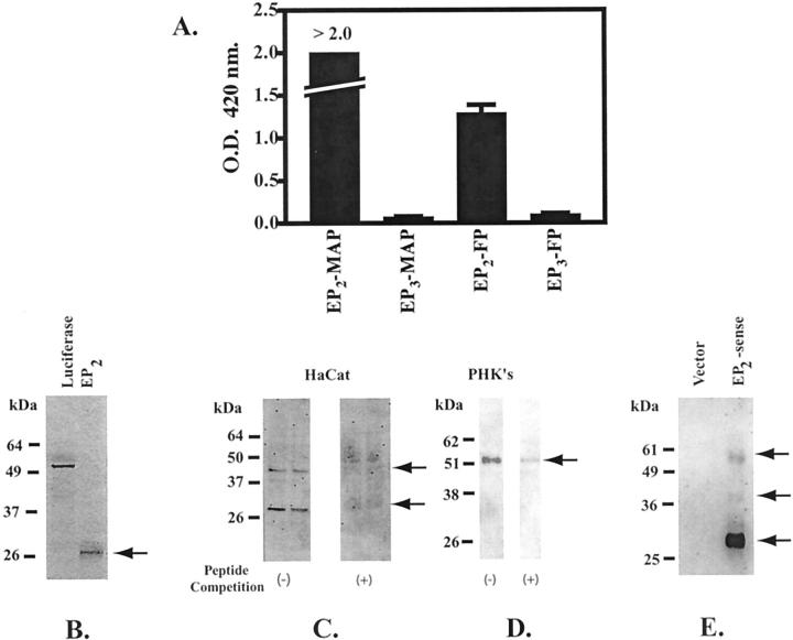
Validation of the anti-human EP2 receptor monoclonal antibody. A: The monoclonal anti-EP2 antibody binds specifically to the immunogenic EP2 amino-terminal peptide sequence. An amino-terminal EP2 peptide coupled to a multiple antigen peptide core (EP2-MAP) was used to elicit the anti-EP2 receptor antibody (clone 2B4). An EIA was used to assess the ability of the anti-EP2 antibody to specifically recognize both the EP2-MAP and the EP2-free peptide sequence (EP2-FP). A MAP peptide coupled to an amino-terminal EP3 peptide (EP3-MAP) and the corresponding free peptide (EP3-FP) are used as negative controls. The results represent the mean and SD for a single EIA done in triplicate. B: Electrophoretic mobility of the EP2 receptor core protein lacking posttranslational modifications. [35S]-Methionine was incorporated into both the EP2 receptor and luciferase by in vitro translation using a rabbit reticulocyte lysate method. Radiolabeled EP2 receptor and luciferase were electrophoresed on a 10% sodium dodecyl sulfate-polyacrylamide gel electrophoresis gel. Bands were detected by autoradiography. C and D: Immunoblot of membrane preparations from HaCat cells (C) and PHKs (D) with the monoclonal anti-hEP2 receptor antibody (clone 2B4). Immunoblots were done in the presence (+) and absence (−) of competing free peptide. E: EP2 receptor expression in membrane preparations from COS-7 cells transiently transfected with an EP2 expression vector. The immunoblot was done using a commercial rabbit polyclonal anti-EP2 receptor antibody (Cayman Chemical, Ann Arbor, MI). The vector control consisted of COS-7 cells transiently transfected with the empty pcDNA3.0 vector.

