
Figure 1.
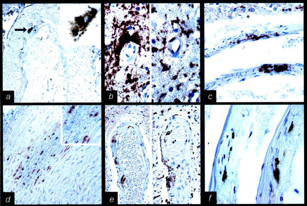
Disease-associated prion protein (PrP) deposits in vessel walls. a: PrP immunoreactivity (IR) (indicated by an arrow) in the media of basilar artery in sporadic Creutzfeldt-Jakob disease (sCJD) (3F4; magnification, ×100; enlarged in right upper corner, magnification, ×750). b: Perivascular PrP IR (left side of picture; 12F10; magnification, ×750) correlates with CD68 IR (right side of picture; magnification, ×750) in variant CJD (vCJD). c: Extensive PrP IR throughout an intracerebral (basal ganglia) vessel wall in vCJD (3F4; magnification, ×500). d: PrP IR in the outer part of the media in extracranial carotid artery in vCJD (6H4; magnification ×200; enlarged in right upper corner, magnification, ×750). e: PrP IR (left side of picture; 3F4; magnification, ×200) in a deep perforating artery in sCJD correlates with CD68 IR (right side of picture; magnification, ×200). f: PrP IR (left side of picture; 3F4; magnification, ×200) in a deep perforating artery in sCJD correlates with HLA-DR IR (right side of picture; magnification, ×200).