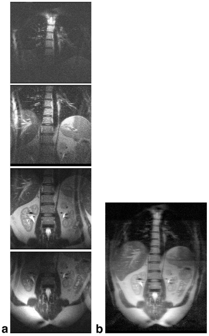
FIG. 7.

a: Reduced-FOV images acquired simultaneously from each row of 8 coils of the 32-element parallel array, with FOV shifted to be centered under that row. Aliasing is evident in the phase-encode direction. b: Combined FASSET image (512 × 1024, FOV 52 cm, imaging time 856 ms), obtained by unwrapping each image in the phase-encode direction, shifting the image by the appropriate amount in the frequency-encode direction, and taking the amplitude-weighted sum of the squares.
