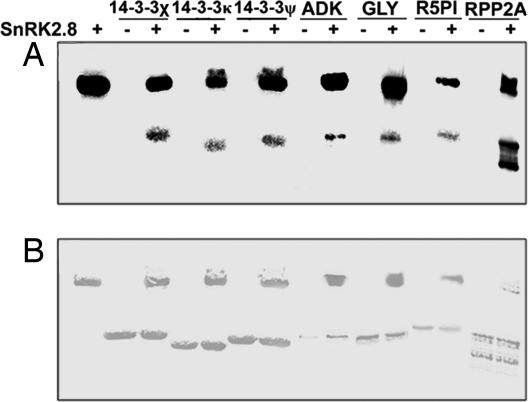
Fig. 4.
In vitro phosphorylation assays conducted by using purified SnRK2.8 protein with three 14-3-3, ADK, glyI (glyoxalase I), R5PI, and RPP2A. (A) Each band indicates a [γ-32P]dATP phosphorylated protein. Lane 1 on the left side contains SnRK2.8 alone. Subsequent lanes contain target proteins that have been incubated with SnRK2.8 (+) or without SnRK2.8 (−). (B) The Coomassie stained gel shows protein loading.

