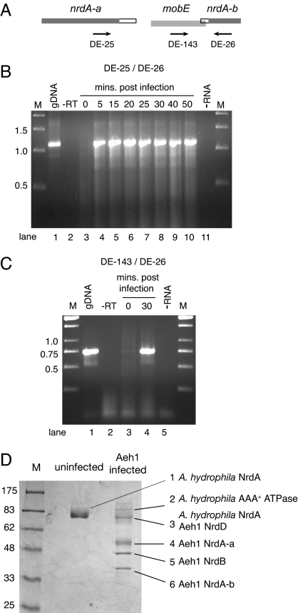
Fig. 2.
The Aeh1 mobE insertion is not a self-splicing intron or intein. (A) Schematic of the Aeh1 mobE insertion indicating the approximate position of primers. (B) RT-PCR with primers DE-25/DE-26. Shown is a 1% agarose gel of aliquots of RT-PCRs using total RNA isolated at the indicated times after Aeh1 infection. gDNA, PCR with Aeh1 genomic DNA; −RT, PCR performed without prior reverse transcriptase step; −RNA, reaction performed without RNA; M, molecular weight markers with sizes indicated in kilobases. (C) RT-PCR with primers DE-143/DE-26 and labeled as in B. (D) Purification of phage Aeh1-encoded RNR proteins from infected A. hydrophila cell extracts by dATP-Sepharose chromatography. Shown is a Coomassie-stained 10% SDS/PAGE gel of a dATP-Sepharose purification using uninfected or Aeh1-infected A. hydrophila cell extracts. Bands corresponding to proteins identified by mass spectrometry are numbered. M, molecular mass marker with sizes indicated in kilodaltons.

