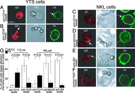
Fig. 3.
LFA-1 cross-linking initiates F-actin polymerization, whereas CD28, NKG2D, or 2B4 cross-linking polarize the MTOC. (A–F) YTS cells (A and B) and NKL cells (C–F) were coated with biotinylated anti-CD18 (the β2 subunit of LFA-1) mAb (A and C), anti-CD28 mAb (B), anti-NKG2D mAb (D), anti-2B4 mAb (E), or anti-class I ME1 mAb as control (F) and then mixed with streptavidin-coated polystyrene beads. After 0.5–2 h at 37°C, the cells were fixed and permeabilized for the intracellular staining of F-actin (green) and MT (red). (D) The percentage of conjugates showing F-actin polymerization at the bead-cell contact site and of MTOC polarization toward the bead-cell contact site (arrows) are shown. P values were calculated after examining >50 images for each experiment. (G) Quantitation and statistical analysis.

