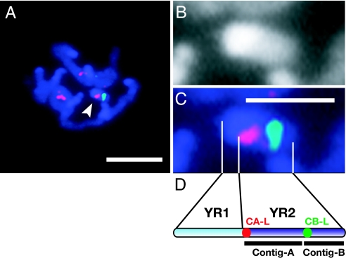
Fig. 1.
Ordering of contig A and contig B on the M. polymorpha Y chromosome. (A) Detection of the left terminal regions of contig A (CA-L, red) and contig B (CB-L, green) on DAPI-stained prometaphase chromosomes (blue). The Y chromosome is indicated by an arrowhead. (B and C) Magnification of DAPI-stained and FISH images of the Y chromosome, respectively. (D) Schematic alignment of contig A and contig B on YR2. The positions of CA-L and CB-L are indicated by red and green dots, respectively. (Scale bars: 5 μm, A; 2 μm, C.)

